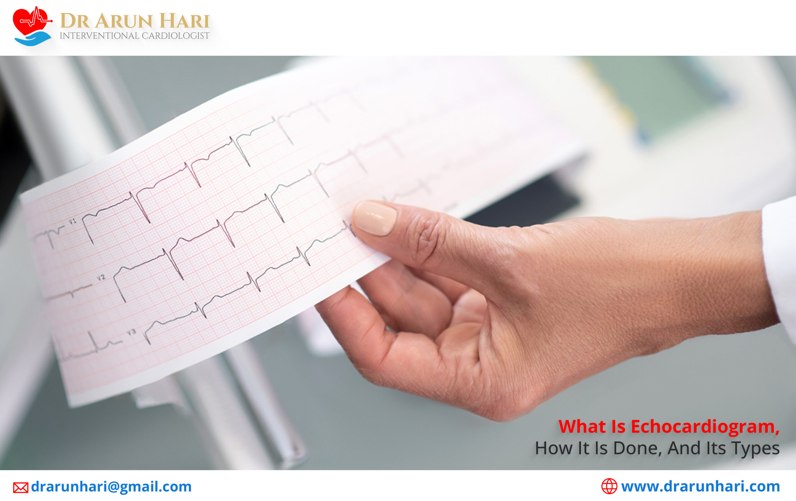
How Echocardiogram (ECG) Is Done & Its Types?
An echocardiogram is a test that uses ultrasound waves to create a moving image of your heart. The image is produced by a machine that sends the waves through a wand held over your chest. The waves bounce off your heart and are transformed into electrical signals that create the image. The test generally takes about 30 minutes on an outpatient basis and is done to evaluate your heart for the following:
- To check for any abnormalities with your heart chambers or heart valves.
- To check if your heart is the reason behind your severe symptoms of shortness of breath and chest pain.
- To check for any congenital defects of the heart.
How Echocardiogram (ECG) Is Done & Its Types?
#1 – Transthoracic echocardiogram – A transthoracic echocardiogram is a standard way of evaluating your heart fully in which you will be asked to lie on your left side on an exam table. Your left side is the best position for getting a clear image of your heart. Your doctor or a technician will apply gel to your chest and then place the wand (transducer) on your skin. The wand then moves around the chest to take pictures of your heart from different angles. Doctor may ask you to hold your breath or to take a deep breath and hold it for a short time while the machine takes a picture. This helps the machine get a clear picture of your heart. You may feel some pressure on your chest from the wand, but it is not painful. Videotape records the pictures that echocardiogram machine produce. The staff then transfers these to a computer, or printed on film. A cardiologist will interpret the pictures. he then generate a reports. Based on its findings, doctor chalks out a detailed treatment plan that includes medications, lifestyle modifications, and/or surgery.
#2 – Transesophageal Echocardiogram – A transesophageal echocardiogram (TEE) is a type of echocardiogram that is performed by placing a small probe in the patient’s esophagus. This allows the doctor to get a clear view of the heart. This is especially useful in diagnosing certain heart conditions. TEE is generally for patients unable to have a standard echocardiogram. That’s due to poor acoustic window or other factors. It is also useful in patients who are at risk for cardiac complications, such as those with a history of heart attack or stroke. TEE is generally very safe and well tolerated by patients. The most common complication is esophageal irritation, which usually resolves on its own within a few days. In rare cases, TEE can cause more serious complications, such as esophageal perforation or cardiac arrest.
#3 – Doppler Echocardiogram – A Doppler echocardiogram is done to assess the heart’s function and to look for any abnormalities again using the ultrasound probe. When these ultrasound waves bounce off your heart cells and blood vessels, their pitch changes. These are Doppler signals. These are useful in determining the direction & speed of your blood flow. Thus, a Doppler echocardiogram is extremely useful in determining the hearts’ pumping function, heart valve disease, or any blood flow abnormalities. It is a painless and non-invasive test. Doctors usually perform it as an outpatient procedure.
#4 – Stress Echocardiogram – It is another type of echocardiogram especially performed to check for any heart issues that are stress-induced and only happen with any type of physical activity or exercise. Here, your doctor will obtain two sets of ultrasound images immediately before and after the exercise. These are to check for any changes that stress or exercise cause.
Dr. Arun Is an Expert in Treating Any Heart Condition with Great Perfection!
If you have a heart condition that is becoming difficult to exactly diagnose or you simply have symptoms of heart disease and wish further treatment, you should contact an expert interventional cardiologist such as Dr. Arun. He has great experience and success in doing all types of minimally-invasive interventional cardiac procedures with the use of latest technology based tools to achieve the best post surgical outcomes.


